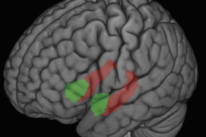
The mere mention of a stressful word like “wheeze” can activate two brain regions in asthmatics during an attack, and this brain activity may be associated with more severe asthma symptoms, according to a study by UW–Madison researchers and collaborators.
The study, which appears in the Proceedings of the National Academy of Sciences (Online, August 29, 2005), reveals a functional link between emotion processing centers in the brain and certain physiological processes relevant to disease.
UW–Madison psychology professor Richard Davidson, an expert on emotions; and UW–Madison medicine professor William Busse, an expert on asthma; are senior co-authors on the study. Melissa Rosenkranz, a graduate student at the UW–Madison Laboratory for Affective Neuroscience, is the lead author.
“While this study was small, it shows how important specific brain circuits can be in modulating inflammation,” says Davidson, director of the affective neuroscience laboratory and the Waisman Laboratory for Functional Brain Imaging and Behavior. “The data suggest potential future targets for the development of drugs and behavioral interventions to control asthma and other stress-responsive disorders.”
Previous studies and clinical evidence have shown that stress and emotional turmoil adversely affect people with inflammatory diseases like asthma. And signs of inflammation have been shown to affect the brain. But until now, nobody knew exactly what brain circuits were involved in these seemingly intertwined emotional and immune events or how the circuits might influence the severity of an acute asthma response.
Researchers used functional magnetic resonance imaging (fMRI) to scan the brains of six mildly asthmatic people who were asked to inhale ragweed or dust-mite extracts. Subjects were then shown three types of words: asthma-related (such as “wheeze”), non-asthma negative (such as “loneliness”) and neutral (such as “curtains”). Shortly after, researchers measured lung function in the subjects as well as molecular signs of inflammation in their sputum.
The fMRI scans revealed that the asthma-related terms stimulated robust responses in two brain regions-the anterior cingulate cortex and the insula-that were strongly correlated with measures of lung function and inflammation. The other types of words were not strongly associated with lung function or inflammation.
The two brain structures are involved in transmitting information about the physiological condition of the body, such as shortness of breath and pain levels, says Davidson, and they have strong connections with other brain structures essential in processing emotional information.
“In asthmatics, the anterior cingulate cortex and the insula may be hyper-responsive to emotional and physiological signals, like inflammation, which may in turn influence the severity of symptoms,” says Davidson.
The researchers suspect that other brain regions may also be involved in the asthma-stress interaction.


