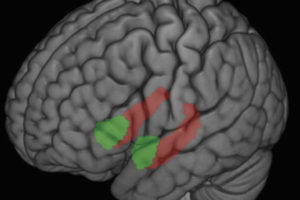
The experiences of millions of people have proved that antidepressants work, but only with the advent of sophisticated imaging technology have scientists begun to learn exactly how the medications affect brain structures and circuits to bring relief from depression.
Researchers at UW–Madison and UW Medical School recently added important new information to the growing body of knowledge. For the first time, they used functional magnetic resonance imaging (fMRI)–technology that provides a view of the brain as it is working–to see what changes occur over time during antidepressant treatment while patients experience negative and positive emotions.
The study appears in the January issue of the American Journal of Psychiatry. UW psychology professor Richard Davidson, psychiatry department chair Ned Kalin, research associate William Irwin and research assistant Michael Anderle were the authors. The researchers found that when they gave the antidepressant venlafaxine (Effexor®) to a small group of clinically depressed patients, the drug produced robust alterations in the anterior cingulate. This area of the brain has to do with focused attention and also becomes activated when people face conflicts. Unexpectedly, the changes were observed in just two weeks.
“Conducting repeated brain scans in these patients allowed us to see for the first time how quickly antidepressants work on brain mechanisms,” said Davidson, who also is director of the W. M. Keck Laboratory for Functional Brain Imaging and Behavior, where imaging for the study took place. He noted that the findings were surprising because patients don’t usually begin noticing mood improvements until after they have been taking antidepressants for three to five weeks.
The researchers also found that while the depressed patients displayed lower overall activity in the anterior cingulate than non-depressed controls, those depressed patients who showed relatively more activity before treatment responded better to the medication than those with lower pre-treatment activity. This kind of information may be extremely useful to clinicians someday, Kalin said.
“We expect that physicians in the future will be able to predict which patients will be the best candidates for antidepressants simply by looking at brain scans that reveal this type of pertinent information,” said Kalin, who also is director of the HealthEmotions Research Institute, where scientists concentrate on uncovering the scientific basis of linkages between emotions and health. One third of all patients treated with antidepressants do not respond to them, and of those that do, only about 50 percent get completely better, he added.
Virtually all previous studies analyzing brain activity in depressed people used PET (positron emission tomography) and SPECT (single photon emission computed tomography) technology. With these imaging systems scientists were not able to obtain pictures with the same resolution as that which is now obtainable with fMRI, which provides a “working snapshot” of the brain.
The Wisconsin team used fMRI’s capability to capture brain activity as it occurred to record subjects’ reactions as they viewed pictures designed to stimulate negative and positive emotions.
“We believe that we can uncover the best indicators of treatment changes when we present research subjects these emotion challenges,” said Davidson. “The pictures activate the individual circuits that underlie different kinds of emotional responses.”
UW emotions researchers have been using fMRIs with emotion-challenging pictures for several years in an effort to understand normal and abnormal brain responses to a range of emotions. They theorize that in depressed people, reactions to negative emotions are similar to, but more exaggerated than, reactions that non-depressed people have, and that the reactions may be more difficult to turn off.
“We all experience some sadness from time to time, but in depression, the responses may be sustained and out of context,” said psychiatrist Kalin.
With the HealthEmotions Research Institute, the Keck Laboratory for Functional Brain Imaging and Behavior and the Laboratory for Affective Neuroscience, UW is home to a critical mass of some of the foremost emotions researchers in the world.


